Showing posts with label Science of Baseball. Show all posts

29 August 2014

Further Discussion of Manny Machado and the Patellofemoral Joint

Wednesday found Orioles third baseman Manny Machado again under the knife, this time to repair a partial tear of his right medial patellofemoral ligament (MPFL); it was performed by Dr. Neal ElAttrache, the same surgeon who repaired his left knee last season. By all reports, the procedure went well and Machado should be able to start the rehabilitation process without any delays. However, news of yet another season ended prematurely by knee injuries for Machado has not only brought skepticism to the rest of season fortunes for the Orioles, but also to the young infielder's chances of a long and productive career. With a 2014 already shortened by an extended rehabilitation from the left MPFL rupture and patellar subluxation suffered in September of 2013 as well as the more recent right knee sprain suffered that evolved into Wednesday's surgery, the revelation of the left MPFL tear casts doubt as to whether he can sustain the physical rigors of the typical MLB season; these injuries also amplify the scuttlebutt on moving Machado to his natural (and more physically demanding) position of shortstop.

For the moment, let's set aside the gloom surrounding the coming years for Machado on the field and take a focused look at the anatomy involved with his knee problems and the surgical procedure he has undergone with Dr. ElAttrache, with the hope that with a better understanding of the medical intricacies of the situation, the doubts and anxiety surrounding Machado's future can be alleviated or at least put into better perspective.

For better or worse, much of the anatomy of the knee joint and the MPFL in particular has been discussed here at Camden Depot; you can find previous articles here and here. Briefly, the MPFL is a ligament that attaches at the femur and patella and helps resist lateral migration of patella, providing a significant amount of the restraining force against the patella dislocating. With the anatomy and mechanisms innate to the MPFL already discussed, let's now shift focus to another crucial piece to the knee joint puzzle and to Machado's health—the anatomy of the trochlea (also called trochlear groove) of the femur and its relationship with the MPFL.

Collectively, this region of interest is called the patellofemoral joint and consists of the patella and its articulation with the trochlea, with the primary role of the patella increasing the mechanical advantage of the quadriceps muscles of the thigh. The stability of this joint is maintained by a complex interaction between soft tissues and bony structures. Stabilizers of the joint are often divided into three groups: active soft tissue stabilizers (the quadriceps), passive soft tissue stabilizers (ligaments) and static stabilizers (the bony anatomy between the patella and trochlea).

With respect to articulations, the patella is covered in cartilage posteriorly that allows for the bone to glide seamlessly along the femur via the trochlea, with seven facets of the patella coming into contact with the femur as the knee flexes and extends. Normal activities typically find five of these seven surface in contact with the femur. The alignment and positioning of the patella is also important and assists in determining the biomechanics of the knee and how the lower legs 'hang', forming the Q-angle, which is an important aspect of the geometry of the knee. The average Q-angle is 14 degrees for males, with anything over 17 degrees considered excessive and called genu valgum—knock-kneed. Smaller Q-angles are thus considered genu varum, or bow-leggedness. Excessive Q-angles can predispose a person to patellar subluxations, such as those suffered by Machado.

Also potentially complicit in patellofemoral instability is the trochlea itself. In the condition trochlear dysplasia, the trochlea is not properly shaped and the patella does not have the normal bony constraints to provide stability. In essence, the trochlear groove is 'flat' and does not exhibit its usual concave shape, giving the articular facets of the patella less surface area to touch, causing a less stable articulation between the two bones. This leads to reliance upon on the MPFL and quadriceps to hold the patella in place, creating a higher than usual propensity for MPFL ruptures and patellar dislocations, due to the decreased strength of the joint. Along with trochlear dysplasia, conditions such as patella alta (a small knee cap) and lateralization of the tibial tuberosity can also affect joint strength. Ligament laxities, such as those seen in Marfan syndrome and Ehlers-Danlos syndrome can also provide patellofemoral instabilities, but are not frequently seen in the older athletic population.

Anatomy of the trochlea and patella. Taken from Practical Orthopaedic Sports Medicine & Arthroscopy 1st Edition, http://www.msdlatinamerica.com
Surgically, both the MPFL repair or reconstruction is a fairly straightforward procedure, with both lending much of the success to the less invasive, arthroscopic approaches that can now be performed. For repairs, tears of the ligament and other soft tissues are typically sutured, often with suture anchors placed in the femur for additional stability and strength, with patellar mobility assessed once sutures and anchors are placed. For a reconstruction, a graft, which is what Machado had previously performed on his other knee, typically the tendon of the semitendinosus muscle is harvested, with tunnels drilled into both the patella and femur for placement of the graft. Passage of the graft and fixation with sutures is also performed, with the native MPFL sutured to the graft and patellar mobility assessed before closure of incisions. It also appears that another procedure on top of either the repair or reconstruction (which is the more likely procedure to have been performed Wednesday) was also undertaken; while no confirmation of the procedure has been found, a common procedure that is often pursued along with the MPFL procedure is a trochleoplasty, which is performed to reshape the trochlear groove, allowing for increased patellofemoral stability.

At the end of all of these procedures, the future still remains slightly fuzzy for Machado. While these operations are necessary and will definitely allow him to return to action for next year, the question remains as to whether he will be the player he was prior to the surgeries. One study in particular looked at outcomes in athletes after MPFL reconstruction found that 100% returned to sports after MPFL reconstruction, 53% returned at equal or higher levels of performance, with 47% returning, but at lower levels of performance. Along with these subjective assessments of return to performance provided by the athletes, knee function was tested using a number of assessments that measured range of motion and pain, with postoperative improvements in these scores predominantly seen. However, there are a couple of caveats to these results, despite their somewhat encouraging tone. First, the athletes themselves were self-evaluating their levels of performance upon return to competition, so biases abound; no sport-specific statistics were used to objectively measure performance returns. Also, the authors included the caveat that these results were found in patients without severe trochlear dysplasia, which is something that could also potentially be ailing Machado.

Overall, youth is on Machado's side, as is the timing of the most recent procedure; the quicker the knee is repaired post injury, the better, and of course, the younger the patient, the more hopeful the medical staff are that a quick, complication-free recovery and rehabilitation will be seen. Using 2014 as a template for Machado's possible return in terms of performance next season, Orioles fans should be confident that his knee woes will be finally nipped in the bud, with his hitting less potentially affected postoperatively than his fielding. For his prowess with the glove, things are a little less certain, with the potential for a deterioration in Machado's lateral quickness a real possibility, especially considering both of his MPFL's are reconstructed. That being said, the future remains promising for Machado remaining in the upper echelon of young baseball talents despite his recent run of knee injuries.


References:

Hamill, J., & Knutzen, K. (2009). Biomechanical basis of human movement. Philadelphia: Wolters Kluwer Health/Lippincott Williams and Wilkins.

Miller, M. (2011). Operative techniques in sports medicine surgery. Philadelphia: Wolters Kluwer Health/Lippincott Williams and Wilkins. 

30 June 2014

Bud Norris' Groin Strain

Bud Norris' quietly successful 2014 campaign was briefly brought to a halt last week when magnetic resonance imaging (MRI) results confirmed a right groin strain injury. Norris, currently enjoying a 7-5 record with 0.7 fWAR and 89 ERA-, left his June 21st start against the New York Yankees after five complete innings pitched after complaining of right leg discomfort; a bullpen session later in the week with continued groin pain prompted the MRI and his current placing on the 15-day disabled list.

Not often associated with disabling injury in baseball, the muscles of the groin—collectively known as the hip adductors (and here on out called 'adductors')—nonetheless can become painful and uncomfortable when strained or torn. A collection of seven muscles that originate from the pelvis that insert into the inner aspect of the femur and tibia,  they are responsible for stabilizing the pelvis and moving the leg towards the midline of the body (adduction), which is where they get their name. Broadly, they are active especially when changing direction during running and in kicking.

Muscles of the groin: adductor brevis, adductor magnus, adductor longus, adductor minimus, pectineus, and gracilis. Obturator externus not shown. Image courtesy of Wikimedia Commons.


With regards to the mechanism of injury, tension is placed on the adductors when they are contracted. When this tension is excessive from repetition or high forces being applied in a short amount of time, one or more of the muscles can tear. Tears can range from a small partial tear with few muscle fibers torn with minimal pain and loss of function, to a complete rupture of one or more muscles, resulting in severe pain and marked loss of function. While there is no universally accepted grading system for muscle strains and tears, the most widely accepted system includes a grade 1 to a grade 4 strain, classified as follows:
  • Grade 1: a small number of muscle fibers are torn; some pain/tenderness but with full function.
  • Grade 2: a moderate number of muscle fibers are torn with moderate loss of function. Tenderness with swelling, possible hematoma and palpable defect.
  • Grade 3: a large number of fibers torn with significant tenderness, swelling, hematoma and partial detraction of muscle. Increased loss of muscle function.
  • Grade 4: all muscle fibers are ruptured resulting in major loss of function, substantial retraction of muscle and corresponding pain, swelling, and hematoma.
The majority of groin strains are grade 2, with the most commonly affected muscle strained being the adductor longus muscle. Factors that could predispose a player to adductor injuries include weakness and/or tightness of the area, perhaps arising from a previous injury in the same leg. For Norris, a right hamstring strain earlier this season, as well as a right hip strain in 2012 and additional right hamstring tightness in 2011 could have possibly put the righthander at greater risk for the adductor strain.

From a pitching biomechanics perspective (and with Norris being a rightander taken into consideration in the following descriptions), the right adductors play a large role in the windup, providing balance at the top of leg kick as well as initiating transfer of power to the upper body by stabilizing the lower extremity and pelvis when the right foot is planted. Through the latter phases of the pitching motion—through stride, arm cocking, arm acceleration, arm deceleration, and follow-through—the right adductors draw the thigh toward midline, pulling the back leg through the delivery and in turn, rotating the hips towards the plate as the pitcher releases the pitch. An electromyography study of the lower extremity in pitching showed that the adductors are the main source of energy that is eventually transferred to the upper extremities, while also contributing to the stabilization of the torso and deceleration of the shoulder and arm in follow-through. As such, insults to the adductors, creating discomfort and a decreased range of motion, can manifest themselves further downstream biomechanically, resulting in shorter stride lengths, leading to poor command of pitches (leaving pitches up in the strike zone) as well as increased stresses put upon the shoulder joint during deceleration and follow-through.

Thankfully for Norris and the Orioles, the groin injury sustained appears to be mild and will resolve with time and the use of one or all of the many physiotheraputic approaches to recovery, including RICE, massage, ultrasound, and stretching. More recent treatment approaches using platelet-rich plasma injections to the offending area are inconclusive, but anecdotally show some acceleration and improvement of the healing process, which can be protracted in lower grade strains. Given the mildness of the injury, Norris should return to the mound without much issue; however, given his history of right upper leg injuries, care will be taken to monitor the righthander's leg health and strength in order to prevent injury re-aggravation.

***

References: Dines, J. S. (2012). Sports medicine of baseball. Philadelphia: Wolters Kluwer Health/Lippincott Williams & Wilkins.


23 March 2014

Science of Baseball: The Most Deceptive Repertoire for a Pitcher, Chris Tillman

Predicting a Baseball's Path
Bahill et al. 2005
American Scientist
Imagine being at the center of the most dramatic moment in baseball. It's the bottom of the ninth inning of the seventh game of the World Series—two outs, the tying run on second, the winning run on first, and you are the batter. Everything depends on you. The trouble is: The most fearsome pitcher in baseball stands on the mound. He has an awesome assortment of pitches: fastball, change-up, curveball, slider and knuckleball. You want any advantage that you can get in predicting where each pitch will go.

With the crowd going wild and sweat pouring from your every pore, you have to concentrate on the ball that is about to be launched in your direction. You must gather as much information about the pitch as quickly as you can in order to make crucial decisions.

As we will show, you get just a few hundreds of milliseconds to figure out what kind of pitch—perhaps traveling at almost 100 miles per hour—is heading toward the plate. In that instant, you must observe the ball's spin and predict how it will move on its way to the plate. It's a daunting computational task. Luckily, we can describe a few clues for you to use. And you will need them soon, because that fearsome pitcher is rocking back on his pivot leg. In a split second, his arm will swing through a great arc and send a baseball hurtling your way.
Usually, we stick to peer-reviewed original research or, when feeling a bit playful, a thesis.  However, the above article comes from a science magazine and is a piece that is too often forgotten.  The article considers pitch movement, how a pitch chances velocity, and other factors a pitcher can use to deceive hitters.  However, my focus will be on the pitch repertoire they suggest.

4S on left, 2S on right
Much of this is already known, but it is good to get actually empirical data on seam flickers with pitches.  Briefly, what they found was that a four seam fastball and a curveball appear the same.  They look white with a slight red tinge.  Meanwhile, a two seam fastball has two lines on it due to the back seam rolling over and over.  This suggests that batters should have more difficulty discerning differences between a four seam fastball and a curveball as opposed to a two seam fastball and a curveball.  As such, we should expect a larger proportion of pitchers utilizing the four seam fastball / curveball combination and that this pitch selection probably results in a more successful pitcher.

A mix of a four seam fastball and a curveball is actually a rather common one and a pairing that is utilized by many successful pitchers (a third of all starting pitchers who had enough innings to qualify for ERA used this approach).  In fact, 69% of pitcher who throw a four seamer more than 40% of the time also utilized a curveball more than 10% of the time.  The list of pitchers who did that is listed below (note: I combined curveball and knuckle curve designations even though the study did not considered recognition issues with knuckle curves. Locke, Minor, Tillman, and Burnett were noted as knuckle curve throwers).


4S CU
Shelby Miller 74 19
Jordan Zimmerman 62 12
Clayton Kershaw 61 13
AJ Griffin 59 16
Jeff Locke 58 19
Matt Harvey 58 13
Mike Minor 57 14
Lance Lynn 56 10
Jose Fernandez 53 34
Jose Quintana 53 19
John Lackey 52 10
Chris Tillman 50 17
Stephen Strasburg 49 23
Julio Teheran 47 13
AJ Burnett 46 35
Felix Doubront 46 14
Eric Stults 45 11
Justin Verlander 44 14
Far fewer starters lean on two seamers, which might be telling in and of itself.  Where the 40% or more four seam use had a group of 26 pitchers, the two seam 40% or more use has a group of twelve (15% of qualified SP).  Only Kris Medlen (44% 2S, 18% CU), Joe Saunders (42% 2S, 11% CU), and Bronzon Arroyo (41% 2S, 12% CU) utilize the curveball to any significant extent.  With such a small sample size, it is difficult to tell whether the group mark of 25% is significantly less than the 69% displayed in the 4S group using their curveballs, but it certainly looks like there is a difference between those populations.

Moving forward, there is a great analytical opportunity I hope to be able to investigate further.  We should be able to take a look at optically similar projectiles and compare that to the flight path in a ball at the last point of batter reaction, which is about a third of the way to the plate.  I would think that the more similar in position a pitch is that looks like each other, the more deceptive it will be.  Likewise, there is probably some sweetspot where dissimilar pitches that take the same path will be as confusing as similar looking projectiles with more different paths.

16 March 2014

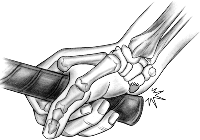
Science of Baseball: Hook of Hamate Fracture in Competitive Baseball Players

Hook of Hamate Fractures in Competitive Baseball Players
Bachoura et al. 2013
Hand

Background

Baseball players are susceptible to a number of specific upper extremity injuries secondary to batting, pitching, or fielding. Fractures of the hook of hamate have been known to occur in batters. The purpose of this study is to present our experience with the surgical management of hook of hamate fractures and their short-term impact on the playing capability of competitive baseball players.

Methods

A retrospective chart review was performed on patients with hook of hamate fractures between the years 2000 and 2012. The inclusion criteria were (1) hook of hamate fracture, (2) competitive baseball players, and (3) surgical treatment of the injury. Patient demographics, mechanism of injury, surgical treatment, and outcome were collected from the medical records. Information on return to play was collected from the Internet when applicable.

Results

There were seven male patients that underwent eight procedures. The mechanism of injury was attributed to batting in six cases and rogue pitches in two cases. All surgeries consisted of hamate hook excision and ulnar tunnel decompression. One patient had concomitant carpal tunnel release. The median time between surgery and return to play was 5.7 weeks (range, 4.3 to 10.4 weeks).

Conclusions

The mechanism of hook of hamate fractures in baseball players is predictable, most often developing secondary to repetitive swinging. This injury may occur at all levels of competition. Ulnar tunnel decompression with hook of hamate excision provides good outcomes, with minimal complications and early return to play.
The above study looked at hook of hamate fractures in seven competitive players.  This is the injury that Nick Markakis suffered in May of 2012.  In the study, they looked at seven players ranging from 19 to 26 years old whose level of play ranged from high school to MLB.  One player suffered two breaks that required surgery each time.  Of these total eight incidents, six were the product of swinging while two happened to being hit by a pitch.  Typically treatment involves excision of the hook of hamate and surgically decompression the ulnar tunnel from around the nerve.  All players returned to action within 30 games or were ready for the start of the next season.  Insufficient data was collected to measure impact on power.

----

In general, the injury is often associated with a quick recovery time (about 5 weeks), but players experiencing lingering weakness with respect to generating power in baseball.  The idea being that the hand itself takes time to be able to firmly grasp the handle.  As far as I am aware, there is no comprehensive study of major leaguers who have had the surgery.  There are decent studies looking at amateurs that found similar levels of performance at the end of the rehabilitation time.

Below is a comparison of Markakis' performance before and after his horn of hamate break in 2012 as well as his performance in 2013.


AVG OBP SLG ISO
2012 Before 256 333 452 196

After 335 390 489 154
2013
271 329 356 85
As you can see, his power collapse in 2013 and it did not do that after the hamate break, which suggests all that you have read about that break impacting his power is likely inaccurate.  What is more likely to have impacted Nick was when C.C. Sabathia broke his thumb with a fastball, ending Markakis' season in September.

09 March 2014

Science of Baseball: Effect of Playing Surface on Batted Ball Velocity

Effects of Surface Conditions on Baseball Playing Surface Pace
Brosnan et al. 2011
Journal of Testing and Evaluation, Vol. 39, No. 3
The speed at which a baseball travels after impact with a playing surface has been referred to as playing surface pace. Little information is available regarding the effects of varying construction and maintenance practices on the pace of baseball playing surfaces. Research was conducted to evaluate the effects of construction and maintenance practices on a non-turfed basepath, Kentucky bluegrass (Poa pratensis L.) turf, and six synthetic turf surfaces. Factors evaluated on the non-turfed basepath included soil compaction at installation, surface scarification, and topdressing with a soil conditioner (calcined clay).The effects of cutting height and thatch thickness were evaluated on Kentucky bluegrass, while the effects of simulated traffic and grooming were evaluated on synthetic turf. On the non-turfed basepath, increasing soil compaction yielded increases in surface pace. Calcined clay topdressing and increasing scarification depth did not affect surface pace. On Kentucky bluegrass, varying cutting height and thatch thickness levels had no effect on surface pace. On synthetic turf, increases in simulated traffic resulted in slight increases in pace. Surface pace measurements on synthetic turf were less variable than those made on natural turfgrass.The results indicate that the pace of commonly used baseball playing surfaces is not easily altered with minimally invasive maintenance procedures and should be addressed at construction or during aggressive renovations.
That abstract might be a bit difficult to parse through.  The link to the pdf of the paper above may also be somewhat problematic.  Really, the take home of this research was that the study evaluated many ways in which the velocity of a 100 mph baseball could differ.  They evaluated three different kinds of turf: Kentucky blue grass, synthetic turfs, and turf-less plots.  None, in this series of experiments, appeared to significantly impact the mean surface pace of the ball.  Surface pace refers to the ratios of the velocity of the ball after striking the ground to the velocity of the ball before striking the ground.  As expected, energy is lost by striking the ground, so all speeds are lower leaving the strike area than they are entering the strike area.  Anyway, ground cover appeared to have no impact on velocity loss.

Grass thickness was also evaluated and yielded no significantly different results.  The heights of the grass evaluated in this study also did not impact velocity loss.  One can imagine though that a wider variety of grass height would eventually produce significant differences, but those differences may only be real at extremes unlikely to be seen at an actual field.  Surface scarification and application of clay top dressing appeared to not affect things either.

Of the variables they did explore, only surface compaction significantly affected velocity loss.  It is difficult to figure out the degree to which this is true because they apparently only repeated the experiment twice, which prevents many different types of statistical evaluations.


Surface Pace
Compaction T1 T2
High 0.543 0.589
Medium 0.525 0.573
Low 0.442 0.527
What the above data suggests is that turf managers would have a hard time affecting how a ball travels.  In other words, the only meaningful way to impact a batted ball on the bounce would be at the time of construction of the field or a rather intensive reconditioning of the playing field.  Certainly some variables were not explored in this paper.  For instance, long grass that is wetted might actually significantly impact surface pace, but that was not explored in this paper.  Additionally, stories we have all heard back in the day about rather clever groundskeepers may have been possible then and not now due to more stringent expectations about playing field conditions.

02 March 2014

Science of Baseball: Vision Training May Improve Performance Dramatically

Science of Baseball is a series that has been on hiatus for a long while.  For the most part, it ended when I ceased to have access to journal articles about a year after my doctorate was finished.  However, I have been able to procure some studies from other sources.  As such, we will try to make this a feature on Sundays to peruse as you take in some afternoon baseball.  Enjoy.

-----

Improved Vision and On-Field Performance in Baseball Through Perceptual Learning
Deveau et al. 2014
Current Biology
Our visual abilities profoundly impact performance on an enormous range of tasks. Numerous studies examine mechanisms that can improve vision. One limitation of published studies is that learning effects often fail to transfer beyond the trained task or to real world conditions. Here we report the results of a novel integrative perceptual learning program that combines multiple perceptual learning approaches: training with a diverse set of stimuli, optimized stimulus presentation, multisensory facilitation, and consistently reinforcing training stimuli, with the goal to generalize benefits to real world tasks. We applied this training program to the University of California Riverside (UCR) Baseball Team and assessed benefits using standard eye-charts and batting statistics. Trained players showed improved vision after training, had decreased strike-outs, and created more runs; and even accounting for maturational gains, these additional runs may have led to an additional four to five team wins. These results demonstrate real world transferable benefits of a vision-training program based on perceptual learning principles.
Snellen Eye Chart
The basic idea presented here is that there may be some elements of baseball that could be enhanced with training regimens that have been shown to be useful in other contexts.  Researchers wound up taking 19 players on University of California - Riverside's (DI) team and putting them through 25 minutes sessions to develop their visual perception and response.  These software sessions included a variety of different exercises that are supposed to enhanced an individual's ability to improve contrast (which can help distinguish seams on a baseball, perhaps) as well as peripheral vision (which can help with head placement when batting).

In laboratory testing, they found that the players that underwent the training improved in two ways.  Their ability to read the Snellen Eye Chart improved significantly.  The individuals also manged to improve their ability to detect contrasts in a well lit environment.  In real life, they found the team exhibiting a roughly four percent decrease in strikeouts, which was significant against the performance of the rest of the league.  Additionally, the team improved by over 40 runs over what the researchers expected to the team to accomplish.  From there, they used the well known Pythagorean Win Expectation to conclude that the team improved by about four games.

Of course, improvement could have resulted from other factors that were unknown or untested by the researchers, so this should not be taken as a conclusive study.  However, it is a remarkable use of technology and certainly a direction to investigate in the future.


16 December 2013

Head Cases: Concussions, Collisions, and Chronic Traumatic Encephalopathy at the Winter Meetings

Despite a quiet Winter Meetings in terms of trade and free agent acquisitions for the Baltimore Orioles, it was a productive one for baseball overall with word that Major League Baseball is working to eliminate home plate collisions no later by 2015, with some believing the rule change will be in place in time for the 2014 season.

With the new rules change being discussed, catchers will not be allowed to block home plate off from oncoming runners, and runners no longer allowed to target catchers, inspiring contact and a collision. If such events were to occur, they will be subject to disciplinary action; umpires will be able to review calls when there are questions of how a collision was provoked and whom by, the catcher or runner.

A source of contention for several years already, many have taken to the internet and other media sources to vocalize their displeasure with the vanquishing of home plate collisions and its effect on the tradition and culture of baseball. In spite of these few dissenters of the proposed changes, the change has been welcome by most, in particular, catchers, with the 'safety first' approach being taken lauded. A recent article by Amanda Rykoff, which includes quotes from a number of managers who were catchers, goes far in feeling the pulse of how the rule changes are being received, in particular with its role in prevent future injuries, especially concussions. While each of the managers expresses an understanding that not every injury at the plate can be prevented, they do all feel that the rule change can do more good than bad in preventing injuries and potentially prolonging careers.

Here at Camden Depot, we have already taken a cursory look at concussions, both in understanding its pathophysiology, as well as what steps are taken to treat concussions; we also have had a brief look at the player performances pre and post concussion for those who suffered one in 2013, with the results being inconclusive.

Let's now go back to those 20 concussions and look at how they happened. How many happened at the plate, if any? Of those suffering concussions, were they all catchers, or was it a mix of other positions and situations?


From the above chart, we have exactly half of the 2013 concussions happening via a foul tip to the catcher's mask, with the next most common cause of injury coming from being hit by a pitch during an at bat. For collisions you would expect at the plate during a close play -- a player-player collision -- only ten percent, or two in total, arose from this cause. Digging deeper and we find that neither of these two player-player collisions occurred at the plate; one came from outfielders colliding, while the other occurred when the baserunner hit his head on a middle infielder's knee trying to break up a play at second base.

Here is a further breakdown of 2013 concussions by position and course of injury:

Position N = Concussion Source (N)
Catcher 11 Foul tip (10), Hit by pitch (1)
Infield 5 Hit by pitch (3), player-player collision (1), player-turf collision (1)
Outfield 3 Player-turf collision (2), Player-player collision (1)
Pitcher 1 Batted ball

Focusing on catchers, we see those aforementioned foul tip inspired concussions, along with one hit by pitch during an at bat -- for those curious, that is Max Stassi of the Houston Astros. Looking across all positions, we only see two player-player collisions, none occurring at the plate.

Overall, we see an interesting disconnect in the 2013 data -- while preventing home plate collisions should absolutely be pursued from the catcher's health perspective, we don't see any tangible instances that could have been prevented were the rules change in place last season. In fact, a vast number of the concussions we see are simply unfortunate happenstance -- the foul tip, a pitch that gets away from a pitcher, even a slight misstep, causing a loss of balance -- and not the result of a premeditated, hard nosed play. That being said, safety is of the utmost importance and any way that injuries can be minimized must be pursued, neurological injury or otherwise, even if our selection biased laden retrospection shows us that concussions at home plate are perhaps better minimized by the improved safety of catcher's equipment. As such, any effort made to improve player health should be pursued, even if for some, the damage is already done.

In a similar vein and a similarly morbid conversation, the Winter Meetings also brought news pertaining to concussions and baseball neuroscience when it was reported that former outfielder Ryan Freel, who took his own life last December due to what many believed was a result of the effects of multiple collisions and concussions sustained, had suffered from chronic traumatic encephalopathy (CTE). Commonly diagnosed in football players post-mortem, it is believed that Freel is the first baseball player to be diagnosed with the still poorly understood disorder; the Boston University Center for the Study of Traumatic Encephalopathy and Sports Legacy Institute released their report that Freel was suffering from Stage II CTE the same day the MLB approved the rule changes regarding home plate collisions. While his Baseball Prospectus player card only reflects two concussions and 65 games lost due to these injuries, it is widely felt that Freel had sustained many more concussions, but had not sought treatment for them, which potentially prompted the cognitive, behavioral, and mood impairments that define CTE.

Briefly, CTE is a progressive neurodegenerative disorder marked by widespread accumulation of hyperphosphorylated tau (p-tau). p-tau is a specialized protein that helps provide structure to the brain; when it becomes hyperphosphorylated, its structure changes, causing neurofibrillary tangles. While most frequently seen in contact sports and in occupations exposed to explosive blast, CTE can occur when exposed to any form of repetitive brain trauma. While concussions are common in CTE, they do not fully explain or bear full responsibility for one developing CTE; being such a 'new' disorder with research only now beginning to be undertaken regarding its pathology, any causal interpretations of CTE should be done cautiously. In short, those with CTE have had a history concussions or other head trauma, but not everyone who has had a concussion will be diagnosed with CTE.

Caveats of correlation implying causation aside, symptomatically, CTE can cause concentration and memory problems as well as disorientation and confusion, dizziness, and headache. Erratic emotions can also transpire, with aggression and psychosis common with CTE. As CTE progresses, behavior issues increase in intensity and erraticism, with aggression and symptoms similar to those of Parkinson's disease. Cognitive abilities decrease, leading to a dementia with more Parkinson's symptoms, including speech and walking abnormalities. There is no cure for CTE and the symptoms cannot be stopped.

With respect to CTE staging, symptoms in stage I chronic traumatic encephalopathy include headache and loss of attention and concentration. Additional symptoms in stage II include depression, explosivity and short-term memory loss, as discussed regarding Freel. For stage III, executive dysfunction and cognitive impairment are present, with dementia, word-finding difficulty and aggression hallmarks of Stage IV CTE.

There is still much research needed to be performed, analyzed, and understood before we can truly understand the extent and long term issues at play in concussive injuries and CTE, in general, and as it relates to baseball injuries. As discussed, the new rules related to home plate collisions are a step in the right direction, but might take years to show any real results in regards to a drop in injuries, concussion or otherwise. If causes for concussion in 2013 are to be used as an example, concussions may not appreciably drop, given many of the concussions arising from unintentional and freak plays. However, with the news of Freel's CTE diagnosis and the knowledge of the man and his style of play potentially leading to his premature demise, anything that the game can do to take better care of their players on the field and 50 years from now should be pursued, given the severity of the symptoms seen in concussions and CTE.

13 April 2013

Adam Jones Blows Bubbles and He Probably Should

Last Friday’s game, Adam Jones dropped a difficult, but catchable, fly ball where he had to turn back and catch the ball running on his glove side.  To make matters visually worse, this happened while he blew a bubble with his gum.  The dropped fly resulted in the fully loaded bases to clear.  It was a pretty aggravating moment.  The Orioles appeared to have gotten out of a tough jam without any of those baserunners scoring.  The fan base response was quick and almost universally in one voice, Adam Jones has to take the game more seriously and stop blowing bubbles.

However, were all of these complaining people wrong?  Yeah, probably.

First, lets think about this in an everyday kind of way.  Yes, our ability to be alert as well as our reaction time as a population is likely not to be similar to athletes.  That said, I think there is still some merit in thinking about this.  What do you do when you are tired at work?  What do you do when you are tired driving in the car?  For those who played some baseball, what did you or others do will you were out in the field?  For most, the answer is a combination of spitting sunflower seeds, drinking coffee/soda/energy drink, smoking a cigarette, partaking in some chaw, and, yes, chewing gum and maybe even blowing bubbles.

Ok, just because people do it, does it actually work?  There are a lot of things people and athletes do that actually provide no measurable benefit.  There are some players who swear that urinating on their hands will help prevent blistering (think about that when you hand one of them a pen to get their autograph).  There is a kernel of truth in that concept.  Urea certainly can help moisturizing your hands and prevent cracking.  Synthetic urea is often an ingredient in hand moisturizer.  However, a meaningful difference exists between urine and commercial moisturizer.  The moisturizer contains ingredients that help saturate the skin with the chemical whereas pure urine does not do it.  You would need to soak your hands in a bucket of it for about 20 minutes to get a similar result.  So, yeah, people do things that result from a misapplication of the truth.

What is the evidence that gum improves alertness or reactions?

Here are a few summaries:
Cognitive Advantages of Chewing Gum (October 2011)
This study randomly assigned different chewing treatments (i.e., gum, sugar-free gum, no chewing) over a population of 159 college students.  What they found was that the students who chewed gum, regardless of sugar content, outperformed non-chewing students in five of six cognitive tests.  The only one they performed worse in was a verbal exercise.

Effects of Chewing Gum on Cognitive Function (February 2010)
This study took 133 people and treated them three ways (i.e., regular gum, fruit-flavored gum, no chewing).  They measured cognitive function in a few ways.  Specifically, they targeted memory, reaction time, memory, selective attention, and sustained attention.  They found that gum did not seem to affect memory.  However, gum chewing participants as a population performed better in cognitive tests and the performance difference became greater as they put them through more difficult problems.

Chewing Gum Moderates Multi-Task Induced Shifts in Stress, Mood, and Alertness (April 2011)
Thirty participants were divided into groups between chewing and non-chewing one day and then flipped the next day.  Among the items they were testing, chewing gum accomplished one thing: improving alertness based on self-assessment.  This finding has been reported in several other papers.

Chewing Gum Alleviates Negative Mood and Reduces Cortisol During Acute Laboratory Psychological Stress (June 2009)
This study used 40 participants similarly to the previous one, different treatments (i.e., chewing gum, not chewing) on different days.  The researchers ran them through through a multi-tasking exercise that has shown to increase stress.  It includes mental arithmetic, the Stroop task, memory search, and visual tracking.  What they found was that if a person was chewing gum, they processed information more quickly and exhibited characteristics that are related to reduced stress.
So what does this all mean?
There is decent evidence that chewing gum can help improve alertness and reduce stress.  This is not a consensus assessment as there is a large, but minority, group of studies that disagree with positive associations with gum chewing.  The primary mode of action for gum being useful is that chewing results in increased blood flow to the brain.  There is also some belief that by actively engaging the mind in low level mental activities that it enables the brain to quickly flip over to stressors of a more primary concern.  In other words, chewing may help the brain from entering a “day dream” mode by keeping it in a more active thought mode.

No, none of this deals with blowing bubbles.  I tried to find some evidence of repetitive mechanical actions improving concentration or reducing stress.  I think I simply am not aware of the proper terminology.  I think we all know people who perform better when doing annoying things like clicking a pen repeatedly.  Feel free to comment with any papers that discuss that.

The take home message is that we have enough evidence that we should not automatically assume that Adam Jones blowing a bubble is detrimental to his performance.  I think it is also fairly obvious that we should refrain from calling on him to no longer chew gum and blow bubbles to reduce the negative outcry because that behavior may well improve his level of play.  In the end, be irritated that he dropped a ball and leave it at that.  You do not need to take the next step and try to explain why it happened.  Simply, we do not know enough.

Let him chew.  Let him blow.  Let him be one of the better players in baseball.

04 April 2013

Science of Baseball: Johan Santana and Anterior Capsule Surgery

by Stuart Wallace


With news of Johan Santana re-injuring the previously repaired anterior capsule of his pitching shoulder and yesterday's successful second surgery, comes an upswell in the curiosity of the injury, what it entails, and what makes it different than some of the more common shoulder injuries befalling players, such as rotator cuff tears and impingements. More commonly seen in traumatic events such as skiing accidents, anterior tears of the shoulder capsule are becoming increasingly diagnosed in baseball as a result of the subacute, microscopic tears of the shoulder capsule that can be sustained with the repetitive, over the head motions native to the game. In spite of the recent advances in orthopedic medicine and imaging technologies, an anterior capsule tear remains indirect and inexact in its diagnosis as a baseball specific injury.


ANATOMY OF THE SHOULDER CAPSULE

Before we begin, for those who would like a primer on the anatomy of the shoulder as it applies to baseball, this article from Baseball Prospectus is a great resource and will provide a firm knowledge base of the relevant shoulder anatomy that will be discussed here.

Briefly, the capsule of the shoulder is a fibrous lining that encompasses the glenohumeral joint and provides additional restraint and stabilization of the joint, keeping the head of the humerus in contact with the glenoid fossa, while also allowing for the wide range of motion allowed by the shoulder joint. It also lends additional support of the glenohumeral joint from the negative pressure environment within the capsule. It attaches laterally to the anatomical neck of the humerus, medially to the glenoid fossa, and superiorly with the attachment of the long head of the biceps, near the root of the coracoid process. The capsule is thickest superiorly and inferiorly and at its thinnest anteriorly; with adduction of the arm, the capsule is loose and lax anteriorly and inferiorly and taut superiorly. 


Illustration courtesy of www.bartleby.com

The capsule is reinforced by a handful of intrinsic ligaments, which themselves are thickenings of the capsule. The anterior portion of the capsule are reinforced by 3 glenohumeral ligaments – the superior, middle, and inferior – which run from supraglenoid tubercle of the scapula to the lesser tubercle and the anatomical neck of the humerus, creating a Z pattern along the capsule. Additionally, the transverse humeral ligament passes from the greater and lesser tubercles of the humerus and provides additional support in keeping the tendon of the long head of the biceps in the bicipital groove, while the coracohumeral ligament strengthens the superior portion of the capsule, and assists in supporting the weight of the arm against gravity at rest and limits inferior and posterior translation of the head of the humerus. The muscles of the rotator cuff reinforce the capsule superiorly, anteriorly, and posteriorly.

 
Lateral cutaway of the capsule. Courtesy of www.seattleclouds.com


INJURY MECHANISMS

Many of the findings on the mechanism of injury leading to anterior capsule tears are similar to those seen with rotator cuff tears. Most commonly, the shoulder capsule is stretched or torn by violent injury such as dislocation or a sudden powerful subluxation. In baseball, the injury more commonly arises from repetitive micro injuries produced from the abduction and external rotation of the shoulder required to perform over the head movements. With abduction, the thinner and weaker anterior capsule  is subjected to multiple iterations of being pulled taut, creating an environment for microscopic tears and stretching of the fibrous capsule to be suffered. With the tear, the normally negative pressure environment within the capsule is lost, thereby reducing stability of the joint, allowing for anterior displacement of the humeral head, and endangering the health of nearby anatomy, in particular, the rotator cuff muscles, as well as the intrinsic ligaments of the capsule.
SYMPTOMS AND DIAGNOSIS OF A CAPSULE TEAR

Differential diagnoses that can cause symptoms similar to a capsule tear are plentiful, and include more  typically seen injuries such as SLAP tears, rotator cuff tears, bursitis, and impingement. Specific to the capsule, a clinician must also rule out adhesive capsulitis, commonly known as 'frozen shoulder', as a cause of the pain.

Much like a rotator cuff tear, the player will complain of pain during throwing, in particular the load and deceleration phases of throwing. This in turn can develop into palpatory tenderness of the shoulder around the insertions of the rotator cuff tendons, as well as the bony landmarks of the glenohumeral joint upon physical examination. Musculoskeletal examinations, including the load and shift test and the apprehension and relocation test can be performed to confirm anterior instability. Magnetic resonance imaging is useful in demonstrating the presence, location, and severity of the capsule tear, usually with the administration of gadolinium contrast necessary.

NON OPERATIVE TREATMENT

Non-surgical approaches to treating capsule tears are predominantly not successful, and demonstrate a high rate of occurrence of re-injury. After a period of rest and the initial management of pain and inflammation has been successful, non-operative methods of treatment focus on the restoration normal range of motion, strength, and mechanics, accomplished through standard physical therapy and baseball specific throwing programs once the player is pain free.

SURGICAL TREATMENT

No matter what surgical technique is used eventually used for a given player, the goal of surgery is to regain stability of the glenohumeral joint through re-establishing the proper amount of tension to the capsule and the restoration of the labrum to its attachment site, should it be torn. While arthroscopic approaches are typically recommended in athletes due to the reduced amount of recovery time and post operative compromises of shoulder range of motion, open arthrotomies are commonly performed with capsule tears, especially when the tear is adjacent to the glenoid cavity versus the humeral head.

To start, the patient is positioned in either the beach chair or lateral decubitus position after anesthesia and intubation have been performed. An exam is performed while the patient is under anesthesia, and is used to assess the degree of joint laxity and confirm preoperative findings and diagnoses. From here, there are slight deviations as to how the surgeon proceeds, depending on whether the repair is open or arthroscopic. In order to expose the capsule in an open procedure – a capsular shift - a number of anatomical elements are reflected away from the surgical site, including the cephalic vein, the deltoid muscle, short flexor tendons, and the subscapularis muscle, which is detached. With the capsule visualized, it is then incised through the tear, creating two flaps, an inferior and superior flap. The rim of the glenoid cavity is then abraded until bleeding to promote healing, and holes are drilled in to the glenoid rim. From here, sutures are placed through these holes and the capsule tissue is sutured to the glenoid rim, with the inferior flap advanced superiorly and the superior flap advanced inferiorly, thereby greatly reducing capsular volume. This procedure not only minimizes laxity of the tissue, but also strengthens the already thin and fragile anterior portions of the capsule by increasing the thickness of the tissue through doubling over the capsule tissue. Multiple sutures can be used to assist in fine tuning the amount and direction of the shift, depending on the severity and location of the tear. Should there be additional pathology with any associated adjacent anatomy, such a a labrum tear, repair of these structures would be performed before the capsular plication and shift is sutured shut. The concepts are similar in an arthroscopic capsular plication technique, with minimal insults to surrounding anatomy, due to smaller incision and surgical sites afforded by arthroscopic technology. With plication, the capsule tear is not extended as it is with an open procedure, and is simply sewn upon itself to the surrounding capsular tissue, again minimizing iatrogenic insults to the anatomy. Before closing the site, passive motion is performed to ensure at least 90 degrees of abduction and 45 degrees of external rotation of the glenohumeral joint are possible.


POST OPERATIVE REHABILITATION

Rehabilitation approaches differ slightly depending on the surgical approach used, but are very similar to those seen with rotator cuff repairs. A thorough and detailed description of the rehabilitation process can be found here. Briefly, range of motion is severely limited for first four weeks following surgery, with only basic daily activities and waist level routines allowed. No over the head movements are permitted for six weeks, and the shoulder is protected through shoulder immobilization and slinging at this time to promote uninterrupted recovery and repair. Hand and elbow exercises to promote full range of motion of these wrist and elbow joints are performed at this stage in the rehabilitation, with some shoulder exercise performed after week 2 of this stage, with everything performed as pain allows, against gravity, and with shoulder rotation kept under 90 degrees. After six weeks, strengthening exercises are started, with slinging and immobilization of the shoulder discontinued. As rehabilitation progresses, strengthening exercises are gradually increased, range of motion exercises are continued, and isokinetic and tubing exercises are introduced around three months post-operation. Assessment of baseball specific function will begin around 3-6 months postoperatively, with throwing programs discussed after about nine months of rehabilitation. With open surgical approaches, this may not happen until later, closer to a year postoperatively. Given the rarity of the anterior capsule repair within baseball circles, these guidelines are still being revised to optimize recovery specific to the procedure.

SUMMARY

There is much to still be learned about the ideal surgical and rehabilitation techniques for an anterior capsule tear in the baseball player. With less than ten players having had confirmed procedures performed for a tear, the verdict is still out as to which methods will provide the ideal mix of total recovery with minimal chance of re-injury, combined with the least amount of postoperative pain and rehabilitation time specific to baseball activities. As imaging techniques improve and awareness of capsule tears within the baseball community increase, improvements will be made not only in the management of tears, but in the prevention of this career threatening injury.